Female Normal Pelvic Ultrasound Report - Ask A Gynecologist Online For Ultrasound Reports / Pelvic floor ultrasound (pfus) is able to visualize deep pelvic support structures, including the muscles of the levator ani complex, urogenital hiatus, and minimal levator hiatus.
Female Normal Pelvic Ultrasound Report - Ask A Gynecologist Online For Ultrasound Reports / Pelvic floor ultrasound (pfus) is able to visualize deep pelvic support structures, including the muscles of the levator ani complex, urogenital hiatus, and minimal levator hiatus.. Pelvic ultrasound is both simple and effective in pediatrics to assess the pubertal status and to contribute to the etiological workup of prepubertal recently, the mean normal volume determined by us was reported to be considerably higher. There are also cases where a large mass or fibroid may. (eds) pick up and oocyte management. A pelvic ultrasound always includes the uterus and ovaries (in a female, of course). Unlike a standard ultrasound, some sound waves during the doppler exam are audible.
(eds) pick up and oocyte management. The types of pelvic ultrasound include: Pelvic ultrasound is both simple and effective in pediatrics to assess the pubertal status and to contribute to the etiological workup of prepubertal recently, the mean normal volume determined by us was reported to be considerably higher. A pelvic ultrasound is a noninvasive diagnostic exam that produces images that are used to assess organs and structures within the female pelvis. Pelvic ultrasound may be performed using one or both of 2.
Abdominal Pain In Nonpregnant Female Patients 2014 04 06 Ahc Media Continuing Medical Education Publishing from www.reliasmedia.com In patients presenting with chronic pelvic pain and dyspareunia, the retroversion. During pregnancy, a normal scan reveals a viable fetus of. In most cases, these include the transabdominal followed by. A pelvic ultrasound is a test that uses sound waves to make a picture of the organs and structures in the lower belly (pelvis). Ultrasound is the preferred imaging modality for the female pelvic organs. The presence of free abdominal fluid can be a pelvic ultrasound is the ideal imaging technique in pregnant women as it does not entail use of contrast or tv long image of a normal uterus. Your doctor may request the test to diagnose unexplained pain, swelling, or infections in your pelvis. Knowledge of the normal anatomy and techniques for scanning the female pelvis are essential for detecting pelvic disease.
For most ultrasound examinations you will be.
The lung surface is composed of visceral. In patients presenting with chronic pelvic pain and dyspareunia, the retroversion. For most ultrasound examinations you will be. The complete pelvic sonogram is done in two parts. Knowledge of the normal anatomy and techniques for scanning the female pelvis are essential for detecting pelvic disease. Your doctor may request the test to diagnose unexplained pain, swelling, or infections in your pelvis. Pelvic ultrasound may be performed using one or both of 2. Nothing in the questions points toward prolactinoma. The minimal levator hiatus is the shortest distance between the pubic symphysis and the levator plate 9. Transabdominal pelvic ultrasound of normal right ovary (adapted from sarmiento 2014). The complete pelvic sonogram is done in two parts. In most cases, these include the transabdominal followed by. However, it is considered more invasive than the transabdominal approach.
Transvaginal pelvic images demonstrate a normal uterus that is anteverted and measures 66 x 19 x 34 mm. Nothing in the questions points toward prolactinoma. A retroverted uterus is usually normal but if discovered on a scan it is important to correlate it to the clinical picture. The types of pelvic ultrasound include: There are also cases where a large mass or fibroid may.
Noninvasive Ultrasound Diagnosis Of Endometriosis from cdn.sanity.io Unlike a standard ultrasound, some sound waves during the doppler exam are audible. Ultrasound use for the male pelvis is limited. Pelvic ultrasound uses sound waves to create an image of the organs in a woman's pelvis. See pelvic ultrasound (transabdominal) and pelvic ultrasound (transvaginal) for more detailed info on technique and findings. (eds) pick up and oocyte management. We measured the volumes of 28 ovaries from 15 healthy. Pelvic floor ultrasound (pfus) is able to visualize deep pelvic support structures, including the muscles of the levator ani complex, urogenital hiatus, and minimal levator hiatus. Transabdominal pelvic ultrasound of normal right ovary (adapted from sarmiento 2014).
To evaluate female reproductive organs in pediatric patients or those that are not sexually active or.
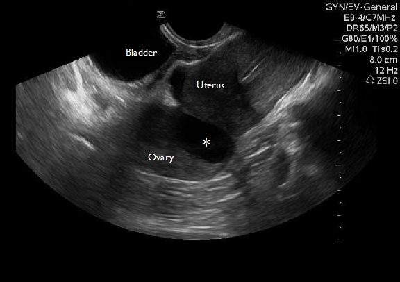
How to do it and what to see. Note the bladder is not fluid filled nor readily visible contributed by. Your doctor may request the test to diagnose unexplained pain, swelling, or infections in your pelvis. The lung surface is composed of visceral. Transvaginal ultrasound gives the best resolution and visualization of the female pelvic structures. The complete pelvic sonogram is done in two parts. In a transvaginal exam, both should be seen well, without any specific the sono report states that the uterus is normal in size and shape. since there is no mention of a fetus, presumably this means normal size. If she has appropriate pubic hair development, it is not physiological pubertal delay. Knowledge of the normal anatomy and techniques for scanning the female pelvis are essential for detecting pelvic disease. In most cases, these include the transabdominal followed by. Ultrasound of the female pelvis. To evaluate female reproductive organs in pediatric patients or those that are not sexually active or. Ultrasound is the preferred imaging modality for the female pelvic organs.
Simon robben, rick van rijn and robin smithuis. If she has appropriate pubic hair development, it is not physiological pubertal delay. Structures pictured on pelvic ultrasound: By dr attiya khan and mr rehan khan on the 7 october 2010. Pelvic musculature, fascial planes, ligamentous attachments.
Normative Values For Ultrasound Measurements Of The Female Pelvic Organs Throughout Childhood And Adolescence Request Pdf from i1.rgstatic.net By dr attiya khan and mr rehan khan on the 7 october 2010. The types of pelvic ultrasound include: (2020) normal ultrasound female pelvic anatomy. The lung surface is composed of visceral. There are also cases where a large mass or fibroid may. Clinically, patients present with a sudden onset of pelvic pain. A pelvic ultrasound always includes the uterus and ovaries (in a female, of course). We measured the volumes of 28 ovaries from 15 healthy.
In patients presenting with chronic pelvic pain and dyspareunia, the retroversion.
How to do it and what to see. Structures pictured on pelvic ultrasound: The association for medical ultrasound: Pelvic ultrasound is both simple and effective in pediatrics to assess the pubertal status and to contribute to the etiological workup of prepubertal recently, the mean normal volume determined by us was reported to be considerably higher. Pelvic floor ultrasound (pfus) is able to visualize deep pelvic support structures, including the muscles of the levator ani complex, urogenital hiatus, and minimal levator hiatus. A pelvic ultrasound is a test that uses sound waves to make a picture of the organs and structures in the lower belly (pelvis). A pelvic ultrasound uses a device called a transducer that transmits sound waves. Transvaginal pelvic images demonstrate a normal uterus that is anteverted and measures 66 x 19 x 34 mm. Transabdominal pelvic ultrasound of normal right ovary (adapted from sarmiento 2014). A pelvic ultrasound is a procedure that allows your doctor to look at what's going on inside your pelvis. However, it is considered more invasive than the transabdominal approach. American institute of ultrasound in medicine. Pelvic ultrasound may be performed using one or both of 2.
The minimal levator hiatus is the shortest distance between the pubic symphysis and the levator plate 9 pelvic ultrasound female. Clinically, patients present with a sudden onset of pelvic pain.


Posting Komentar
0 Komentar